| Hierarchical Morphology-Guided Tooth Instance Segmentation from CBCT Images | |||
| Zhiming Cui1,6,7, Bojun Zhang2, Chunfeng Lian3, Changjian Li4, Lei Yang1, Wenping Wang1,5, Ming Zhu2, Dinggang Shen6,7 |
1The University of Hong Kong, 2Shanghai Ninth People’s Hospital, 3Xi’an Jiaotong University, 4 University College London, 5Texas A&M University, 6 ShanghaiTech University, 7 Shanghai United Imaging Intelligence Co., Ltd. |
||
| International Conference on Information Processing in Medical Imaging (IPMI), 2021 | |||
| Oral Presentation |
|||
 |
|||
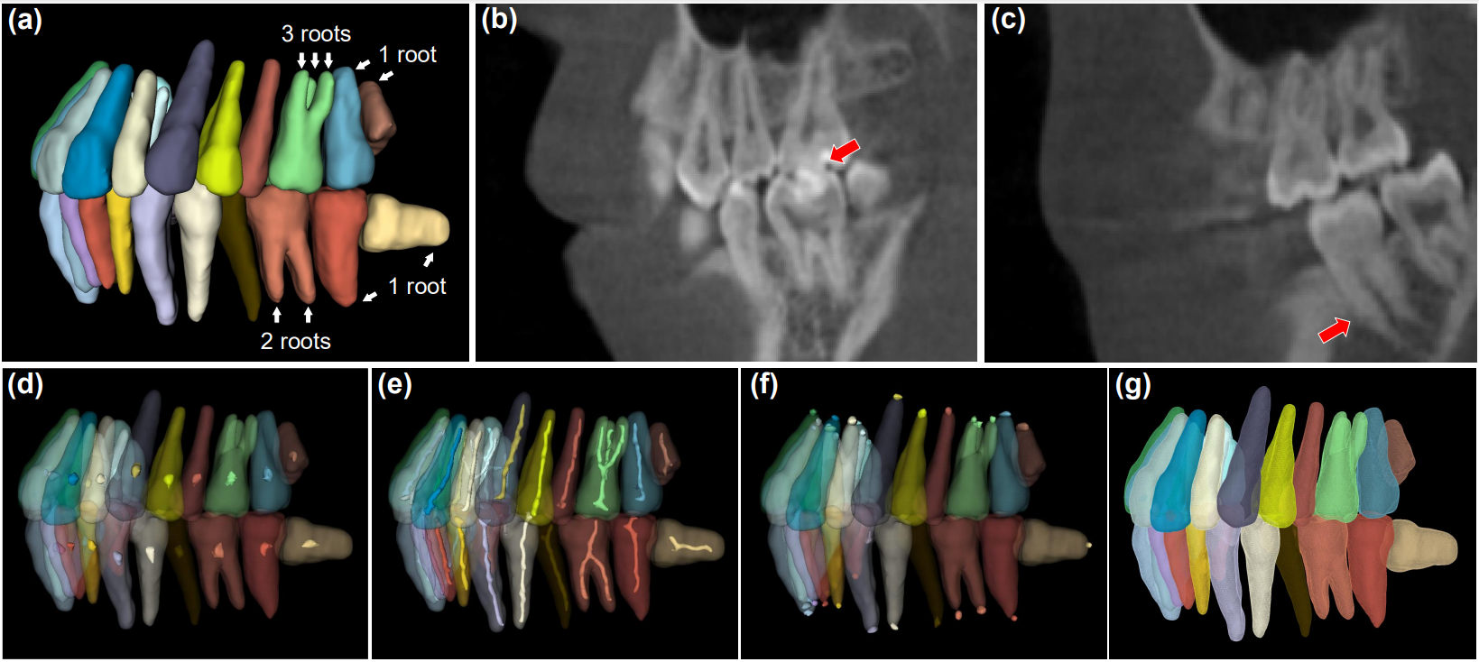
| Fig. 1. The first row shows three typical examples, including (a) teeth with large shape variations; (b) touching boundaries of maxillary and mandibular teeth during close bite; (c) blurred signals between tooth roots and the surrounding alveolar bones. The second row illustrates different components of the hierarchical morphological representations, including the points (i.e., the tooth centroid (d) and the root landmarks at root apices (f)), the tooth skeleton (e), and the tooth boundary surface (g). | |||
| Abstract | |||
| Automatic and accurate segmentation of individual teeth, i.e., tooth instance segmentation, from CBCT images is an essential step for computer-aided dentistry. Previous works typically overlooked rich morphological features of teeth, such as tooth root apices, critical for successful treatment outcomes. This paper presents a two-stage learningbased framework that explicitly leverages the comprehensive geometric guidance provided by a hierarchical tooth morphological representation for tooth instance segmentation. Given a 3D input CBCT image, our method first learns to extract the tooth centroids and skeletons for identifying each tooth’s rough position and topological structures, respectively. Based on the outputs of the first step, a multi-task learning mechanism is further designed to estimate each tooth’s volumetric mask by simultaneously regressing boundary and root apices as auxiliary tasks. Extensive evaluations, ablation studies, and comparisons with existing methods show that our approach achieved state-of-the-art segmentation performance, especially around the challenging dental parts (i.e., tooth roots and boundaries). These results suggest the potential applicability of our framework in real-world clinical scenarios. | |||
|
|||
|
|
|||
| Network Overview | |||
 |
|||
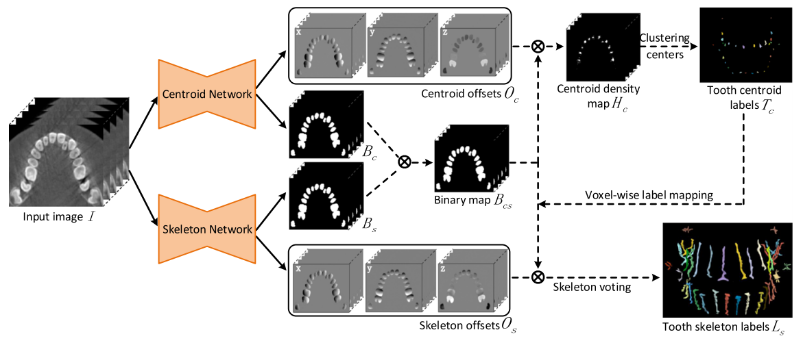
| Fig. 2. The pipeline of our 1st-stage network for tooth skeleton extraction. The CBCT scan is first fed into both the centroid network and the skeleton network to generate the offsets and binary maps, respectively. Then, the tooth centroids and skeletons are detected and predicted by the later steps (dotted lines). | |||
 |
|||
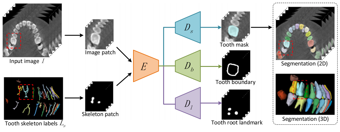
| Fig. 3. The pipeline of our 2nd-stage multi-task network for tooth instance segmentation guided by skeletons. The inputs are the cropped input image and skeleton patches cropped from the 3D input image and tooth skeleton label map, and the outputs are the tooth segmentation volume, boundary, and root landmarks. | |||
| Results | |||
| Result Gallery | |||
 |
|||
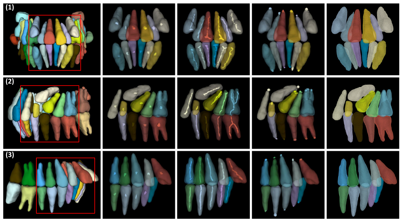
| Fig. 4. Typical results of the hierarchical tooth morphological representation. From left to right: 3D segmentation results, predicted tooth centroids, skeletons, root apices, and boundaries. The last four columns are the partial results of the first column within the red boxes. (Color figure online) | |||
| Comparison | |||
 |
|||
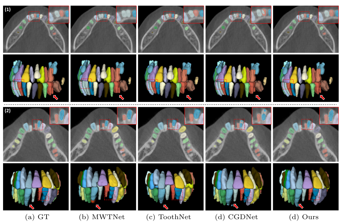
| Fig. 5. The visual comparison of tooth segmentation results by four different methods. Two typical examples are presented, each being shown by two rows with 2D segmentation masks and corresponding 3D reconstruction results, respectively. | |||
| Follow-up Work: | |||
| Zhiming Cui, Yu Fang, Lanzhuju Mei, Bojun Zhang, Bo Yu, Jiameng Liu, Caiwen Jiang, Yuhang Sun, Lei Ma, Jiawei Huang, Yang Liu, Yue Zhao, Chunfeng Lian, Zhongxiang Ding, Min Zhu, and Dinggang Shen. "A fully automatic AI system for tooth and alveolar bone segmentation from cone-beam CT images." Nature communications 13, no. 1 (2022): 2096.[Paper] | |||
|
|
|||
| ©Changjian Li. Last update: June, 2021. |